MIDOG 2021
The first edition: scanner-agnostic mitotic figure detection in human breast cancer, with 200 training cases digitized on four different whole-slide scanners.
MIDOG 2021 is over. If you are interested in joining it's successor, MIDOG 2022, here is a link: https://midog2022.grand-challenge.org
If you are interested in the challenge paper, please find it at: https://www.sciencedirect.com/science/article/abs/pii/S1361841522003279
Motivation
- Mitosis detection is a key component of tumor prognostication for various tumors, including breast cancer.
- Scanning microscopy slides with different scanners leads to a significant visual difference, resulting in a domain shift.
- This domain shift prevents most deep learning models from generalizing to other scanners, leading to strongly reduced performance.
Scope
- Detect mitotic figures (cells undergoing cell division) from histopathology images (object detection).
- You will be provided with images scanned by 4 different scanners, 3 out of which are labeled. In total the set consists of 200 cases of breast cancer.
- Evaluation will be done on four scanners (two new, two are part of the training set) with the F1 score as main metric.
The MIDOG challenge in brief
From the abstract of the challenge report (Aubreville et al., Medical Image Analysis, 2023):
The density of mitotic figures within tumor tissue is known to be highly correlated with tumor proliferation and thus is an important marker in tumor grading. Recognition of mitotic figures by pathologists is known to be subject to a strong inter-rater bias, which limits the prognostic value. State-of-the-art deep learning methods can support the expert in this assessment but are known to strongly deteriorate when applied in a different clinical environment than was used for training. One decisive component in the underlying domain shift has been identified as the variability caused by using different whole slide scanners. The goal of the MICCAI MIDOG 2021 challenge has been to propose and evaluate methods that counter this domain shift and derive scanner-agnostic mitosis detection algorithms. The challenge used a training set of 200 cases, split across four scanning systems. As a test set, an additional 100 cases split across four scanning systems, including two previously unseen scanners, were given. The best approaches performed on an expert level, with the winning algorithm yielding an F_1 score of 0.748 (CI95: 0.704-0.781). In this paper, we evaluate and compare the approaches that were submitted to the challenge and identify methodological factors contributing to better performance.
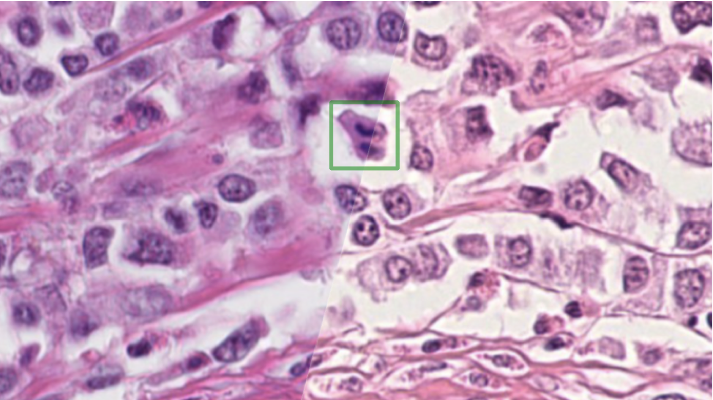
Breast cancer tissue, acquired using a Hamamatsu NanoZoomer XR (left, Scanner A) and a Hamamatsu NanoZoomer S360 (right, Scanner B). Besides a color shift, the depth of field is also affected by using a different scanner, caused by different optical properties of the objective. A mitotic figure in anaphase is indicated by a green box. (Figure 1 of the challenge report)